Home
News: iSeg-2019 is open: https://iseg2019.web.unc.edu/, with more challenging testing subjects from multiple sites.
News: iSeg-2017 journal paper was published in IEEE Transactions on Medical Imaging (download as personal use only), 38 (9), 2219-2230, 2019
If you use the iSeg-2107 challenge data or evaluation framework, please cite the following paper:
 |
|
The first year of life is the most dynamic phase of the postnatal human brain development, along with rapid tissue growth and development of a wide range of cognitive and motor functions. This early period is critical in many neurodevelopmental and neuropsychiatric disorders, such as schizophrenia and autism. More and more attention has been paid to this critical period. For example, Baby Connectome Project (BCP) is recently started and will acquire and release thousands of infant MRI scans, which will greatly prosper the community.
Accurate segmentation of infant brain MR images into white matter (WM), gray matter (GM), and cerebrospinal fluid (CSF) in this critical period is of fundamental importance in studying both normal and abnormal early brain development. Of note, there are three distinct phases in the first-year brain MRI, including (1) infantile phase (≤5 months), (2) isointense phase (6-8 months), and (3) early adult-like phase (≥9 months). In the isointense phase, the intensity range of voxels in GM and WM are largely overlapping (especially in the cortical regions), thus leading to the lowest tissue contrast and creating the most significant challenge for tissue segmentation, in comparison to images acquired at other phases of brain development. For example, Fig. 1 shows longitudinal MR images for an infant scanned every 3 months during the first year, starting from the second week after birth.
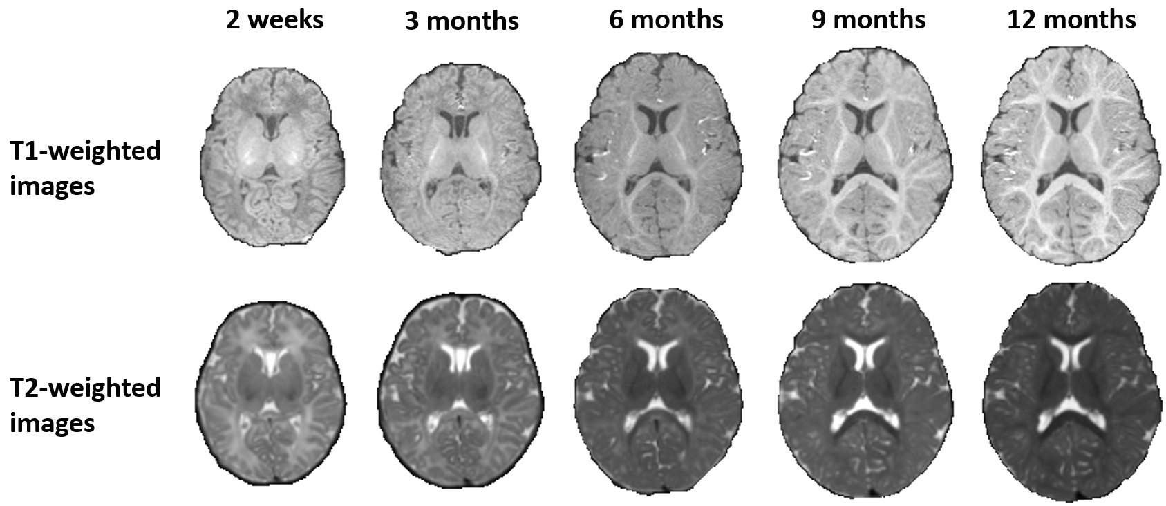
Fig. 1. T1- and T2-weighted MR images of an infant scanned at 2 weeks, 3, 6, 9 and 12 months of age. At around 6 months of age, MR images show the lowest tissue contrast and create the most significant challenge for tissue segmentation.
Many studies have been done on both neonatal and early adult-like brain MRI segmentation. To date, only a few studies focused on the segmentation of 6-month infant brain images [1,2,3] (with the following video showing our previous work, LINKS [1], on segmentation of the challenging 6-month infant brain MRI). In this challenge, researchers are invited to propose and evaluate their automatic algorithms to segment WM, GM and CSF on isointense (6-month) infant brain MRI scans. The aim of this challenge is to promote automatic segmentation algorithms on 6-month infant brain MRI.
The iSeg-2017 will be always open and wating for your submission.
[HD quality movie is available in YouTube]
[1]. Li Wang, Yaozong Gao, Feng Shi, Gang Li, John H. Gilmore, Weili Lin, Dinggang Shen. LINKS: Learning-based multi-source IntegratioN frameworK for Segmentation of infant brain images, Neuroimage, 108, 160-172, 2015.
[2]. Li Wang, Feng Shi, Yaozong Gao, Gang Li, John H. Gilmore, Weili Lin, Dinggang Shen. Integration of Sparse Multi-modality Representation and Anatomical Constraint for Isointense Infant Brain MR Image Segmentation, Neuroimage, 89, 152-164, 2014.
[3]. Wenlu Zhang, Rongjian Li, Houtao Deng, Li Wang, Weili Lin, Shuiwang Ji, Dinggang Shen. Deep Convolutional Neural Networks for Multi-Modality Isointense Infant Brain Image Segmentation, Neuroimage, 108, 214–224, 2015.
![]()